Figure 6.
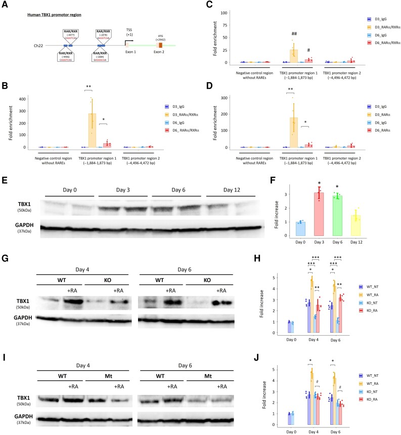
ChIP assays highlight the human-specific binding site of RARα/RXRα complexes on the TBX1 promoter region. (A) Schema showing novel (human-specific) and putative RAR/RXR binding sites (i.e. RARE) on the human TBX1 promoter region. (B) The ChIP assays demonstrated that recruitment of RARα/RXRα complexes onto one of the novel RARE sites of the human TBX1 promoter (−1884–1873 bp) was augmented at Days 3 (D3) and 6 (D6) in CM differentiation of WT hESCs. *P < 0.01 and **P < 0.0001 vs. IgG (negative control). (C) The ChIP assays using STRA6-KO cells demonstrated that RARα/RXRα complexes were also recruited onto the identified RARE site of the human TBX1 promoter (−1884–1873 bp) at Days 3 and 6 in CM differentiation but to a much lesser degree compared with that in WT cells in (B). #P < 0.01 vs. IgG and vs. D6_RARα/RXRα in WT (B). ##P < 0.001 vs. IgG and vs. D3_RARα/RXRα in WT (B). (D) The ChIP assays using only the anti-RARα antibody demonstrated that recruitment of RARα onto the identified RARE site of the human TBX1 promoter (−1884–1873 bp) was augmented at Days 3 and 6 in CM differentiation of WT hESCs again. *P < 0.01 and **P < 0.0001 vs. IgG. (E) Western blotting analysis for expression of TBX1 protein with GAPDH protein (a loading control) in WT cells at Days 0, 3, 6, and 12 in hESC-CM differentiation. (F) Quantitative results in (E). *P < 0.01 vs. Day 0. (G) Comparison of expression of TBX1 protein between WT and STRA6-KO cells at Days 4 and 6 in hESC-CM differentiation with or without treatment with retinoic acid (RA, 0.5 μM) during Days 3–7. GAPDH was used as a loading control. (H) Quantitative results in (G). NT, normal treatment (without adding RA). *P < 0.01 between the NT-administered and RA-co-administered WT cells at Days 4 and 6, respectively. **P < 0.05 between the NT-administered and RA-co-administered STRA6-KO cells at Days 4 and 6, respectively. ***P < 0.01 between WT and STRA6-KO cells under the same treatment conditions at Days 4 and 6, respectively. (I) Comparison of expression of TBX1 protein between WT and TBX1 promoter-mutant (Mt) cells at Days 4 and 6 in hESC-CM differentiation with or without treatment with RA (0.5 μM) during Days 3–7. GAPDH was used as a loading control. (J) Quantitative results in (I). *P < 0.01 between the NT-administered and RA-co-administered WT cells at Days 4 and 6, respectively. #P = not significant between the NT-administered and RA-co-administered TBX1 promoter-mutant cells at Days 4 and 6, respectively. Differences between groups were examined with one-way ANOVA followed by Tukey–Kramer post hoc test.

