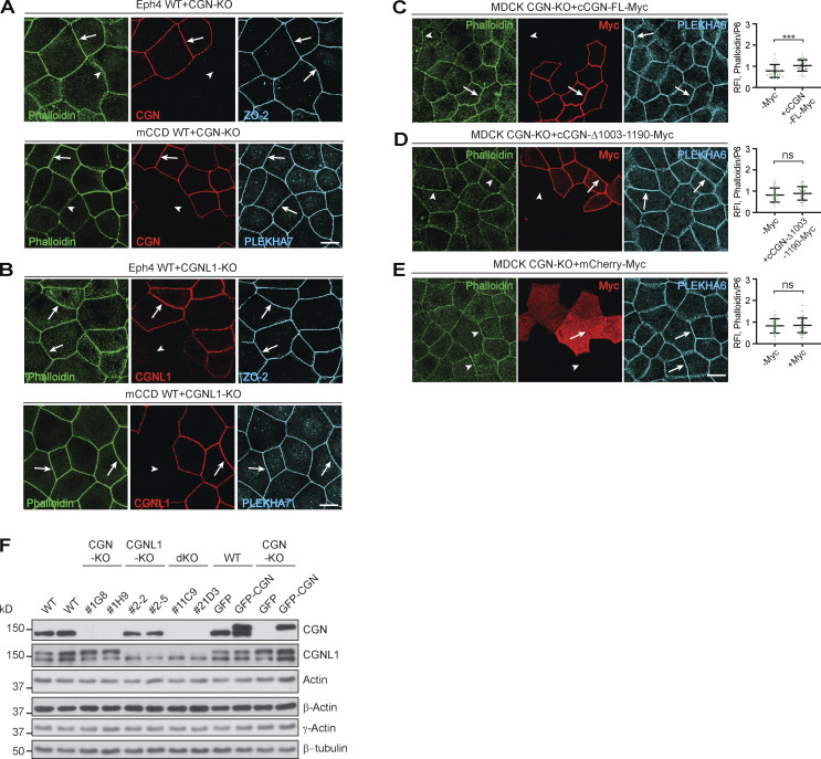
Figure S6.
CGN regulates actin filament organization at tight junctions of mCCD and Eph4 cells. (Related to Fig. 7.) (A–E) IF microscopy analysis of phalloidin labeling either in mixed cultures of either WT + CGN-KO (A) or WT- + CGNL1-KO (B) Eph4 (top panels) and mCCD (bottom panels) cells, or in CGN-KO MDCK cells rescued either with myc-tagged full-length (FL) CGN (C), or C-terminally truncated CGN (D, Δ1003-1190), or mCherry (E, negative control). Either PLEKHA7 or ZO-2 or PLEKHA6 were used as junctional reference marker in mCCD or EpH4 or MDCK cells, and exogenous proteins were detected with antibodies against the myc tag. Arrows = normal labeling and arrowheads = reduced/undetectable cytoplasmic labeling. (F) IB analysis using antibodies against CGN, CGNL1, total actin, β-actin, and γ-actin (β-tubulin as loading control) of lysates from different clonal lines of MDCK cells (WT, CGN-KO, CGNL1-KO, double-KO [dKO]) and from CGN-KO MDCK cells rescued with either GFP-cCGN-myc or GFP-myc. Numbers on the left show migration of prestained markers. The scale bars (normal magnification panels, A–E) represent 10 μm. (C–E) Data in plots are represented as mean ± SD (n = 48 from two independent experiments). Statistical significance was calculated with unpaired Mann–Whitney test (***P < 0.001). RFI, relative fluorescence intensity. Source data are available for this figure: SourceData FS6.

