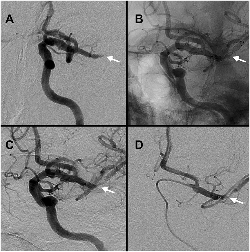
Figure 2.
Case of middle cerebral artery ICAD-LVO on angiography. Panel A: ICAD-LVO tapered occlusion (white arrow). Panels B-C: recanalization with deployed stentriever conforming to the underlying stenosis (white arrow, B unsubtracted, C subtracted). Panel D: post-recanalization angiography confirming ICAD with severe stenosis (black arrow).

