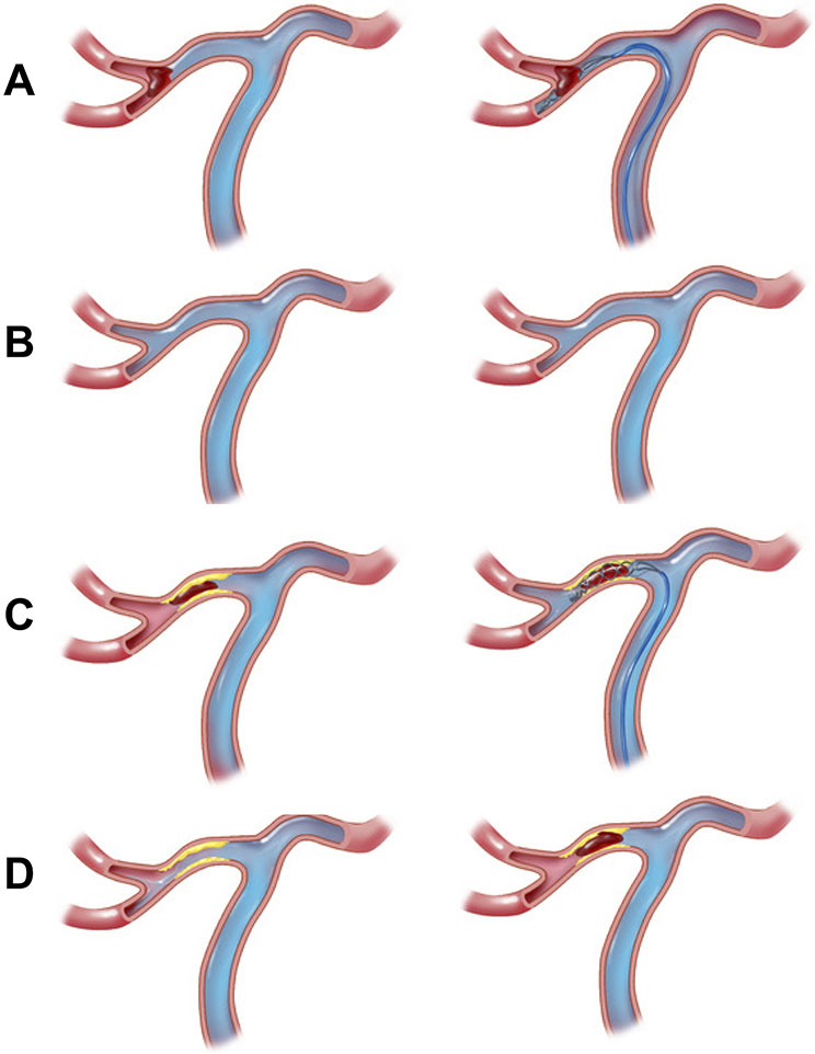
Figure 3.
Middle cerebral artery occlusion angiographic and EVT treatment images. Panels A-B (cardioembolic): a branch segment occlusion characteristic of atrial fibrillation that is successfully recanalized with a stent-retriever on follow-up angiography (panel B, right); Panels C-D (ICAD): a truncal occlusion characteristic of ICAD that is successfully recanalized with a stent-retriever (panel D, left) followed by re-occlusion due to ICAD (panel D, right). Note contrast flow when stentriever is deployed in the inferior division only with embolic LVO (panel A, right) compared to flow in both superior and inferior divisions in ICAD LVO case (panel C, right)

