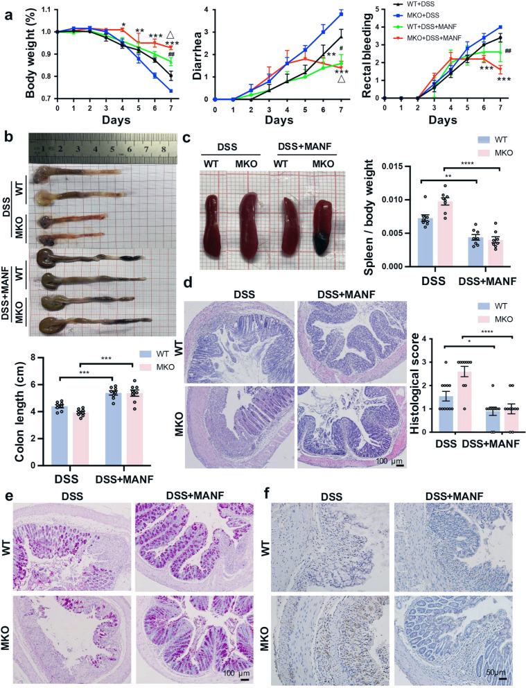
Fig. 4. Exogenous rhMANF alleviates DSS-induced mouse colitis.
WT and MKO mice were injected by tail vein with His-rhMANF (1 mg/kg) from the 4th day of DSS treatment for 3 consecutive days and then were sacrificed. The control group was injected with PBS in the same way. a The time-dependent curves of body weight, diarrhea, and rectal bleeding. Data are collected from three independent experiments with n = 10 mice/group in each experiment and expressed as the mean ± SEM. *P < 0.05, **P < 0.01, ***P < 0.001, DSS+MKO vs DSS+MKO+rhMANF; #P < 0.05, ##P < 0.01, DSS+WT vs DSS+WT+rhMANF; △P < 0.05, DSS+WT+rhMANF vs DSS+MKO+rhMANF. b Gross morphology of colons from WT and MKO mice. Colon length was measured on day 8 (n = 10). c Gross morphology of spleens from WT and MKO mice. Spleen weight was measured at day 8 (n = 10). d Representative H&E staining of colon sections and histology scores of WT and MKO mice. Bars, 100 μm. In panels b and c, data are expressed as the mean ± SEM. *P < 0.05, **P < 0.01, ****P < 0.0001, DSS vs DSS+MANF. e Periodic acid–Schiff staining of colon sections. Bars, 100 μm. f Immunohistochemical staining of CD68 in colon tissues of WT mice and MKO mice (n = 10).

