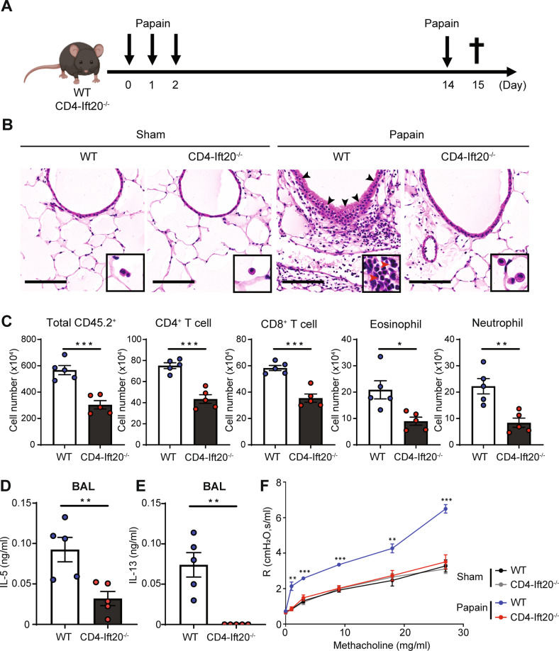
Fig. 2. IFT20 deficiency alleviates protease-induced airway inflammation.
A Overview of the papain-induced airway inflammation model. Mice were sensitized to a protease allergen by administration of 25 μg papain via intranasal injection for three consecutive days. The animals were then intranasally challenged with the same concentration of papain on Day 14 after the first intranasal injection, and lung tissue and bronchoalveolar lavage (BAL) fluid were harvested on Day 15. B Representative hematoxylin and eosin (H&E) staining images of lung tissue from papain-treated and sham control-treated WT and IFT20-KO mice. Black and red arrows indicate goblet cell hyperplasia and eosinophils, respectively. Scale bar, 100 μm. C Total numbers of CD45.2+ immune cells (n = 5), CD4+ T cells (n = 5), CD8+ T cells (n = 5), eosinophils (n = 5), and neutrophils (n = 5) in the lungs of papain-treated WT and IFT20-KO mice. Levels of (D) interleukin (IL)-5 (n = 5) and (E) IL-13 (n = 5) in BAL fluid from papain-treated WT and IFT20-KO mice were measured by enzyme-linked immunosorbent assay (ELISA) (n = 5). F Airway resistance to intratracheal methacholine in WT and IFT20-KO mice following administration of papain (n = 5). The representative images in (B) were obtained from more than two independent experiments. In C–F, the data shown represent more than three independent experiments. The data are shown as the mean ± SEM. Significance was calculated by unpaired Student’s t test. *P < 0.05, **P < 0.01, ***P < 0.001

