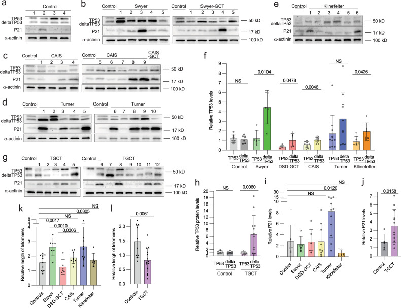
Fig. 5. Expression of TP53 protein in leukocytes of DSD- and TGCT-individuals.
Immunoblotting for TP53 and P21 proteins for controls (a), Swyer and Swyer-GCT (b), CAIS and CAIS-GCT (c), Turner (d) and Klinefelter (e) individuals. (f) Relative quantification full-length (TP53) and depleted isoform (deltaTP53) of TP53 in DSD-groups. Immunoblotting (g) and relative quantification normalized to α-actinin (h) for TGCT-group. Relative quantification of P21 protein in DSD (i) and TGCT (j) groups. Quantification of relative length of telomeres in DSD (k) and TGCT (l) samples.

