
Fig. 1.
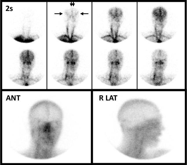
Normal lipophobic perfusion study using 99mTc-DTPA in a young man following a ruptured aneurysm of the left posterior inferior cerebral artery. Eight sequential two-second flow images appear in the upper panel and show excellent visualization of the common carotid arteries as well as the anterior (double vertical arrows) and middle (horizontal arrows) cerebral arteries, forming a “trident” appearance, which indicates presence of intracranial perfusion. The activity progresses to a transient blush of activity within the parenchyma followed by visualization of the intracranial venous sinuses on the final flow images. On immediate static images (lower row) in anterior (ANT) and right lateral (R LAT) projections, activity is noted in the venous sinuses but there is no localization within the brain parenchyma—an expected and normal finding when imaging lipophobic RPs. This Figure was originally published by author L. S. Z. in the Journal of Nuclear Medicine (Zuckier LS. Radionuclide evaluation of brain death in the post-McMath era. J Nucl Med 2016; 57: 1560–8, © SNMMI) and is reproduced in accordance with applicable Authors’s Permission to Reprint (https://jnm.snmjournals.org/page/permissions [accessed October 2022])