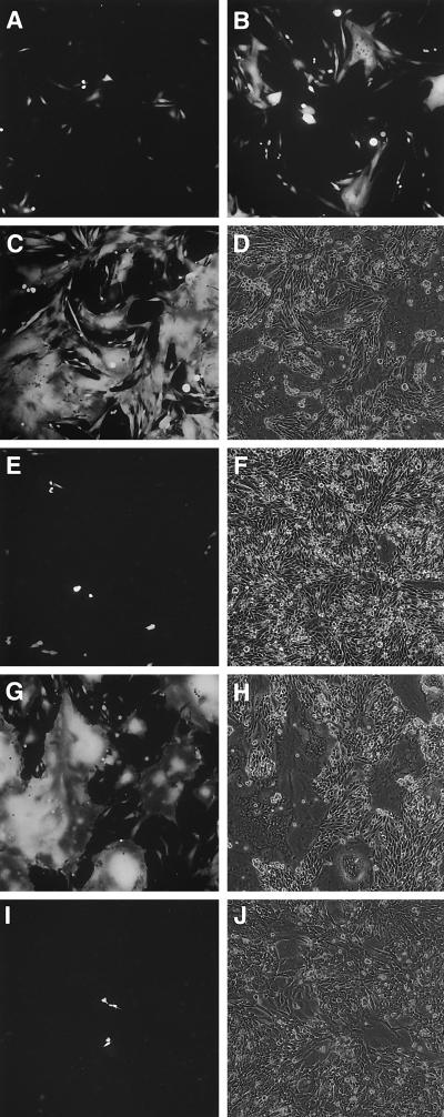
FIG. 3.
Infection of CHO-hEGFr cells (A to F), CHO-hEGFr.tr cells (G and H), and CHO cells (I and J) with MVgreen-H/XhEGF (A to D and G to J) or MV (E and F). Cells were infected at a MOI of 1 and were monitored by fluorescent or phase-contrast microscopy 24 (A), 48 (B), or 72 (C to J) h after infection.

