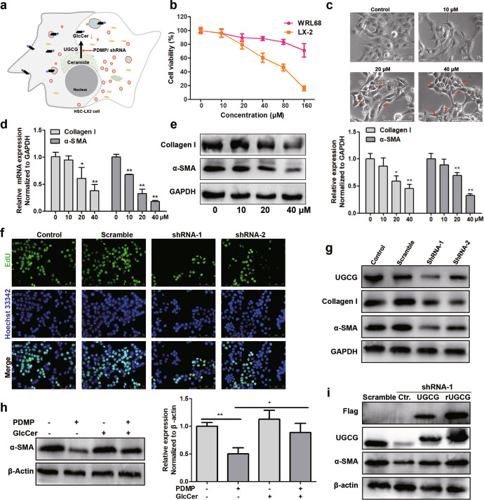
Fig. 2. The inhibition of UGCG suppresses the activation of HSCs in vitro.
a Schematic diagram of strategy for intervention of UGCG. b MTT assay of PDMP-treated WRL68 or LX2 cells for 48 h. c Cell morphology assessment, red arrows indicate the representative intracellular vesicles. d The mRNA levels of Collagen I and α-SMA after treatment of cells with different PDMP concentrations, as detected by qPCR. e The protein levels of Collagen I and α-SMA were detected by Western blot. f The effect of UGCG knockdown on the proliferation of LX2 cells, as detected by an EdU kit. g The expression levels of UGCG, Collagen I and α-SMA were evaluated by Western blot. h Effect of exogenous GlcCer (2 μM) on the expression of α-SMA. i UGCG knockdown cells were rescued with UGCG or rUGCG. The experiments were repeated three times, and statistical significance was determined by a t-test. *P < 0.05, **P < 0.01.

