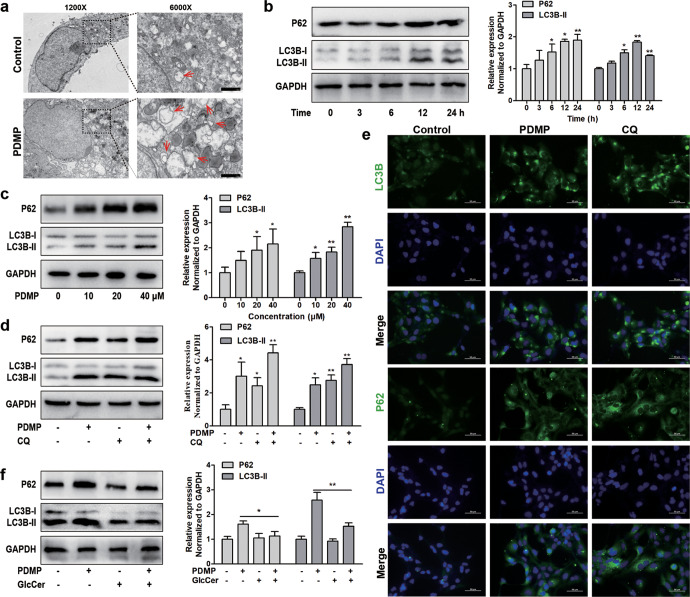
Fig. 3. The inhibition of UGCG blocks autophagy flow.
a Human HSC-LX2 cells were pretreated with 40 μM PDMP for 12 h and microstructure of cell was analyed by transmission electron microscopy, red arrows indicated autophage (Scale bar, 1 μm). b The expression levels of LC3B and P62 were evaluated in different time periods by Western blot. c The expression levels of LC3B and P62 were evaluated by Western blot analysis in LX2 cells treated with different concentrations of PDMP. d The expression levels of LC3B and P62 were evaluated by Western blot analysis in LX2 cells treated with PDMP (40 µM), CQ (20 µM), or CQ (20 µM) + PDMP (40 µM). e The expression levels of LC3B and P62 were evaluated by immunofluorescence analysis in LX2 cells treated with PDMP (40 µM) or CQ (20 µM). f The expression levels of LC3B and P62 were evaluated by Western blot analysis in LX2 cells treated with PDMP (40 µM), GlcCer (2 µM), or GlcCer (2 µM) + PDMP (40 µM). The experiments were repeated three times, and statistical significance was determined by a t-test. *P < 0.05, **P < 0.01.

