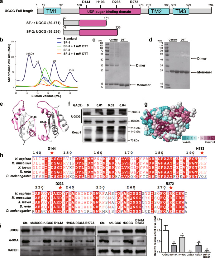
Fig. 5. Study on protein structure and activity of UGCG.
a Domain organization of UGCG, with boundaries indicated. The transmembrane domain (TM) and UDPG binding domain are colored in blue and red. b The gel filtration running trace of SF-1 and SF-2 with a Superdex 200 increase column, superimposed on the chromatogram of standard protein markers. c and d SDS-PAGE analysis of purified SF-1 and SF-2 with or without 1 mM DTT by coomassie blue staining. e The predicted dimer of UGCG. f UGCG protein in LX2 cells was crosslinked with glutaraldehyde, and Keap1 was used as positive control. g Sequence conservation analysis of UGCG (the conservations scales were shown in lower right). h Multiple sequence alignment was performed for Homo sapiens (H. sapiens), Mus musculus (M. musculus), Danio rerio (D. rerio) and Drosophila melanogaster (D. melanogaster) for UDPG binding domain of UGCG. i Rescue effect of different mutants of UGCG was evaluated by Western blot. The experiments were repeated three times, and statistical significance was determined by a t-test. *P < 0.05, **P < 0.01.

