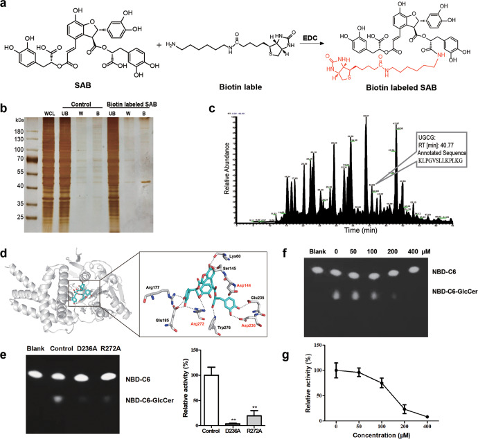
Fig. 7. Salvianolic acid B targets UGCG.
a The chemical structural formula of biotin-labeled SAB. b SAB-binding proteins were visualized by silver staining. WCL: whole cell lysates, UB: the unbound proteins, W: the washing proteins, B: SAB-binding proteins. c Total Ion Current (TIC) chromatograms from 4 to 65 min. d Molecular docking of SAB with UGCG, hydrogen bonds formed within 3.5 Å. e The activity of UGCG mutations was detected by thin layer chromatography. f and g The activity of UGCG was detected by thin layer chromatography. The experiments were repeated three times, and statistical significance was determined by a t-test. *P < 0.05, **P < 0.01.

