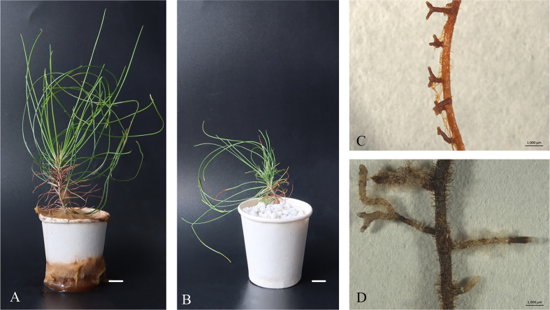
Figure 5.
Morphology of P. massoniana ectomycorrhiza in regenerated plantlets. (A) Plantlets after inoculation with the ectomycorrhizal fungus Pisolithus orientalis. Scale bar = 1.0 cm. (B) Uninoculated plantlets. Scale bar = 1.0 cm. (C) Details of a root with dichotomously branched, ectomycorrhiza-like structures viewed under a Leica MZ16 stereomicroscope. Scale bar = 0.1 cm. (D) Non-inoculated root. Scale bar = 0.1 cm.

