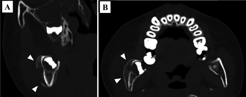
Fig. 3.
(A) Computed tomography coronal-section image. A radiograph revealed the presence of a fistula in the buccal cortical bone of the tooth germ of the third molar (arrowheads), and periosteal hyperostosis was observed on the outside. (B) Computed tomography sagittal plane image. Periosteal thickening of the right angle of the mandible was observed (arrowheads).

