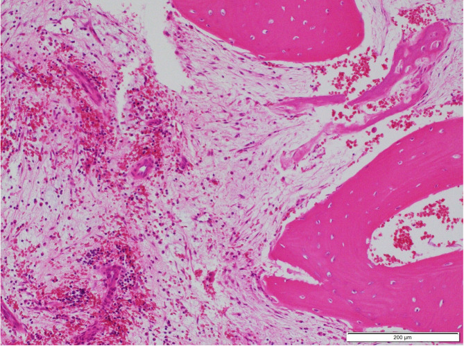
Fig. 7.
Histopathological images. Osteocytes were observed in the lacuna, and fibrosis and hemorrhage between the bone trabeculae. Further, mild lymphocyte infiltration and chronic osteomyelitis occurred and bony trabeculae of the reactive bone arranged perpendicular to the bone surface. However, there was no jaw osteonecrosis. Hematological and eosin staining. Bar = 200 μm.

