
FIGURE 4.
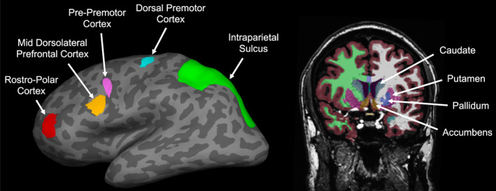
Region of interest (ROI) locations in a representative participant. Left: Cortical ROIs shown on the inflated left hemisphere (dark gray = sulci, light gray = gyri). Nomenclature of the frontal ROIs as in Badre et al. (2010). Right: Subcortical ROIs including the dorsal striatum (caudate and putamen, shown in teal and purple, respectively), ventral striatum (accumbens, shown in light brown), and pallidum (shown in blue). Other colors correspond to other anatomical segmentations by Freesurfer (white and green = white matter in the left and right hemispheres, respectively, covered by gray matter shown in dark brown).