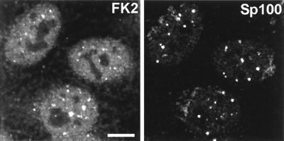
FIG. 1.
Distribution of conjugated ubiquitin in uninfected HEp-2 cells and its presence in some ND10. HEp-2 cells were simultaneously stained for conjugated ubiquitin with MAb FK2 (left panel) and for the ND10 component Sp100 with rabbit serum SpGH (right panel). A proportion of the major ND10 foci contain local accumulations of conjugated ubiquitin. Bar, 10 μm. The magnifications of most panels in other figures are similar, but in all cases the average width of a HEp-2 cell nucleus is 10 to 15 μm. The original colored images of this and all other multichannel figure panels may be viewed at http://www.vir.gla.ac.uk/staff/everettrd/JVI832.shtml.

