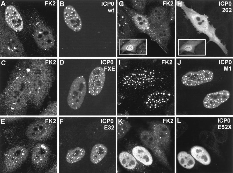
FIG. 2.
Expression of ICP0 in transfected cells leads to accumulation of colocalizing conjugated ubiquitin. HEp-2 cells were transfected with plasmids expressing wild-type and mutant forms of ICP0 and then stained to detect ICP0 with rabbit serum r191 (r95 for plasmids 110262 and E52X) and conjugated ubiquitin with MAb FK2. Each pair of panels shows the FK2 (left) and r191 (right) staining of the same field of view. (A and B) Wild-type ICP0. Note the difference in FK2 staining between the transfected (upper left) and untransfected (lower two) cells. Superimposition of the two images in the original data showed precise colocalization of the FK2 and ICP0 foci. (C and D) ICP0 RING finger deletion mutant FXE. (E and F) Exon 2 insertion mutant E32. The transfected cells are clearly distinguishable from the untransfected cells in the FK2 staining, but the result is not as striking as with the wild-type protein. (G and H) Exon 1 and exon 2 truncation mutant 110262 (ICP0R). The transfected cells have clearly altered FK2 staining patterns that in some cells had a punctate character (main panel) but was sometimes diffuse (inset). The 110262 staining pattern was generally diffuse throughout the whole cell. (I and J) USP7 binding-deficient point mutant M1. (K and L) C-terminal truncation mutant E52X. Although this mutant has a generally diffuse nuclear staining pattern, the transfected cells were easily distinguishable by examining the FK2 staining.

