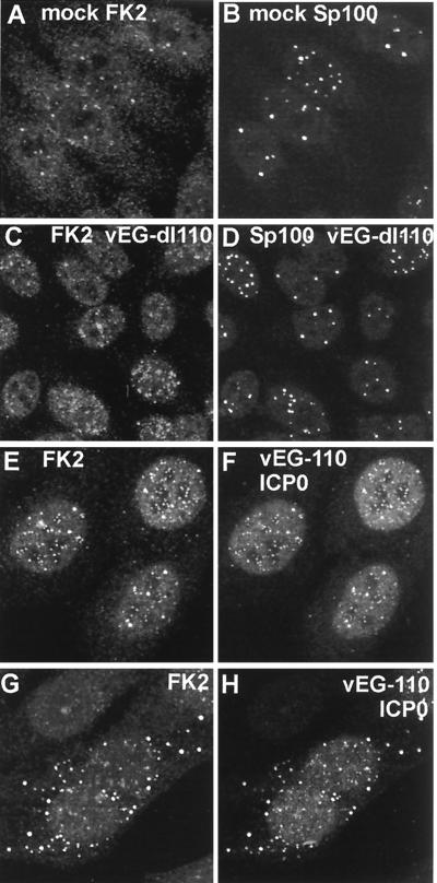
FIG. 5.
Effect of HSV-1 infection on the distribution of conjugated ubiquitin in HEp-2 cells at 2 h postinfection. Each pair of images shows the same field of view with the conjugated ubiquitin (FK2) staining on the left (A, C, E, G) and either ND10 protein Sp100 (B and D) or ICP0 (F and H) staining on the right. (A and B) The distribution of conjugated ubiquitin in uninfected cells. A few foci are embedded in a generally diffuse distribution throughout the cell. Superimposition of the two images showed that some of the conjugated ubiquitin foci colocalize with ND10 (not shown). (C and D) Infection with ICP0 null mutant vEG-dl110 results in increased numbers of conjugated ubiquitin foci in the nucleus. The original triple-labeled image with the EGFP signal to identify infected cells is available on http://www.vir.gla.ac.uk/staff/everettrd/JVI832.shtml. (E and F) Infection with virus vEG-110 expressing wild-type ICP0 fused to EGFP results in induction of conjugated ubiquitin foci that colocalize with ICP0. Infected cells were easily detected on the basis of the FK2 staining alone. (G and H) When ICP0 accumulates in foci in the cytoplasm, it induces the formation of colocalizing conjugated ubiquitin here also.

