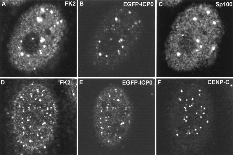
FIG. 6.
Triple colocalization of ICP0 and conjugated ubiquitin at ND10 and interphase centromeres in the early stages of infection. HEp-2 cells were infected with vEG-110 and stained for FK2 and either Sp100 or CENP-C as indicated. ICP0 was detected by the linked EGFP signal. All the major Sp100 foci (C) contain ICP0 (B), and all the ICP0 foci (B) react with MAb FK2 (A). In a certain proportion of cells, ICP0 also colocalizes with interphase centromeres, as shown here (D through F). The major ICP0 foci (E) are likely to be at ND10, but many of the minor foci are at centromeres (F). Of the 27 centromere foci visible in this confocal optical slice (F), at least 11 both contain ICP0 (E) and react with MAb FK2 (D). Because of the difficulty of reproducing the correct color balance of triple-colored images in printed journals, the single-channel data are shown here in greyscale. The original triple-colored images may be viewed at http://www.vir.gla.ac.uk/staff/everettrd/JVI832.shtml.

