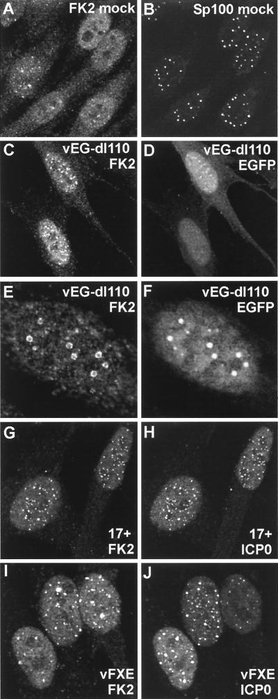
FIG. 9.
Effect of expression of mutant ICP0 proteins on the distribution of conjugated ubiquitin during HSV-1 infection of HFL cells. The panel pairs show on the left the conjugated ubiquitin staining and on the right either Sp100 staining (B), EGFP (D and F), or EGFP-linked ICP0 (H and J). (A and B) The relationship between the distributions of conjugated ubiquitin and ND10 is similar to that in HEp-2 cells. (C and D) Infection of HFL cells by ICP0 null mutant vEG-dl110 for 2 h has a more marked effect on conjugated ubiquitin than in HEp-2 cells, with more prominent induction of nuclear foci, which are commonly associated with ND10 (not shown). EGFP itself forms foci in some cells, which can be associated with the conjugated ubiquitin foci. (E and F) At later times of infection, conjugated ubiquitin forms ringlike structures in some cells, and these frequently contain internal EGFP in vEG-dl110 infections. The faint clouds of EGFP in panel F correspond to replication compartments, which are generally not associated with the conjugated ubiquitin ring structures (which can occur in all mutant ICP0 virus infections but never in wild-type infections). (G to J) At 3 h postadsorption, EGFP-linked wild-type ICP0 is strongly associated with conjugated ubiquitin foci, but the RING finger deletion mutant FXE shows a variable degree of colocalization.

