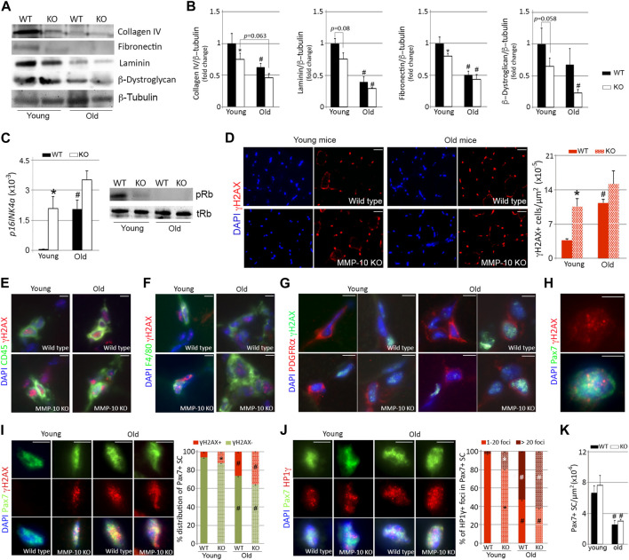
FIGURE 1.
MMP-10 loss is associated to muscular aging and it confers aging features to the satellite cells. Representative western blots of the TA muscles from wild type and MMP-10 KO mice at 2 (young) or 18 (old) months of age (A), using antibodies to proteins indicated, and quantification of ECM protein levels (B). p16INK4a gene and pRb protein (C) expression in muscles of wild type and mutant mice at different ages. Representative muscle tissue sections of wild type and MMP-10 KO mice at 2 and 18 months of age immunostained for γH2AX (D), CD45/γH2AX (E), F4/80/γH2AX (F), PDGRFα/γH2AX (G), Pax7/γH2AX (H, I) and Pax7/HP1γ (J). Graphs show total number of γH2AX+ cells in the muscles (D), percentage of Pax7+ satellite cells containing positive γH2AX (I) and positive HP1γ foci (J). Total number of Pax7+ satellite cells per μm2 in the TA muscles of wild type and KO mice at different ages (K). DAPI was used to identify all nuclei. Scale bar: 40 μm. All numerical data were expressed as the mean ± SEM from at least three mice per genotype and age, including two independent tissue sections per mice. * designates significance between wild type and MMP-10 KO mice at the same age, while # defines significance between young and old mice of same origin, where * and # p < 0.05. mo, months; KO, MMP-10 knockout mice; WT, wild type mice; TA, tibialis anterior.

