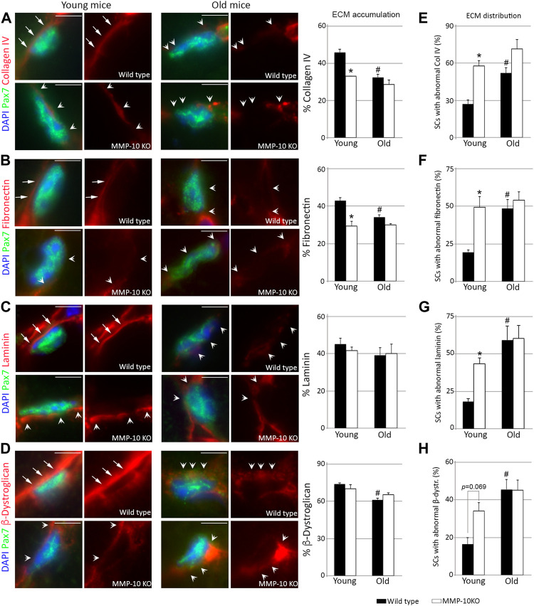
FIGURE 2.
Loss of MMP-10 modifies the satellite cell niche ECM. Representative TA muscle sections of wild type and KO mice at 2 and 18 months of age co-immunostained for Pax7 and collagen IV (A), fibronectin (B), laminin (C) and β-dystroglican (D). All MEC protein images were taken at the same exposure time. Arrows identified the continuity of the ECM proteins in the Pax7+ satellite cells from young wild type mice, while arrowheads highlight an abnormal ECM, with disruptions or rare accumulation of the proteins. DAPI was used to identify all nuclei. Scale bar: 5 μm. Graphs show the average accumulation of each protein in the niche ECM (A–D) and the percentage of Pax7+ satellite cells with abnormal protein distribution in the niche ECM (E–H). All values were expressed as the mean ± SEM from three animals per condition, analyzing two independent tissue sections per mice. * designates significance between wild type and MMP-10 KO mice at the same age, while # defines significance between young and old mice of same origin (p < 0.05). KO, MMP-10 knockout mice; WT, wild type mice; SCs, satellite cells; β-dystr, β-dystroglican.

