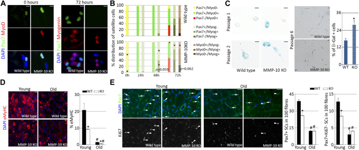
FIGURE 3.
Satellite cells from young MMP-10 KO mice function as cells from aged mice. Representative images of single EDL myofibres isolated from young MMP-10 KO and wild type mice stained for Pax7 and MyoD or Myogenin, immediately after isolation or after 72 h in mitogen-rich media (A). Distribution of the satellite cells according to the expression of different pairs of myogenic transcription factors, immediately after isolation and after 24, 48, and 72 h in mitogen-rich media (B). Satellite cells from young KO and wild type mice stained for β-galactosidase after consecutive passages and percentage of senescent cells at passage 6 (C). Representative images of the TA muscles of wild type and KO mice at different ages stained for eMyHC (D), a protein expressed in fetal and newly regenerating fibers (Sacco et al., 2010), and Pax7/Ki67 (E) after two episodes of injury. Arrows and arrowheads indicate Pax7+Ki67+ and Pax7+Ki67- satellite cells, respectively. DAPI was used to identify all nuclei. Scale bar: 5 μm (A, C), 40 μm (D, E). Percentage of regenerating fibers (D) and number of Pax7+ and Pax7+Ki67- satellite cells in 100 fibers (E) in young and old wild type and KO mice 7 days after last episode of damage. Values are presented as the average of at least three independent experiments (mean ± SEM). * and # designate significance (p < 0.05) between wild type and KO mice at the same age or between adult and aged mice of same strain, respectively. KO, MMP-10 knockout mice; WT, wild type mice; β-Gal, β-galactosidase; eMyHC, embryonic Myosin; SCs, satellite cells; TA, tibialis anterior; EDL, extensor digitorum longus.

