FIGURE 4.
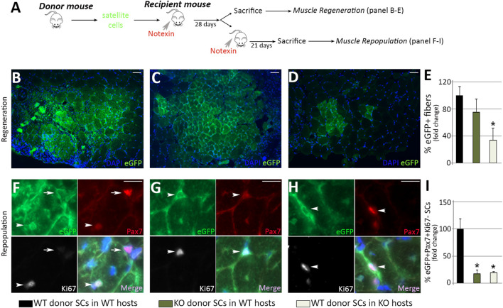
Loss of MMP-10 induces intrinsic and extrinsic changes in the satellite cells at a young age. Experimental schematic outlining the transplantation of satellite cells isolated from donor mice into the TA muscles of pre-injured recipient mice (A). Representative images of recipient muscles stained for GFP (B–D) and percentage of new GFP+ regenerating fibers of donor origin at 28 days after cell delivery (E). TA sections of recipient mice stained for GFP, Pax7 and Ki67 (F–H) and total number of GFP+Pax7+Ki67- quiescent satellite cells of donor origin (I) 3 weeks after inducing damage in recipients. Arrows and arrowheads identify Pax7+Ki67- and Pax7+Ki67+ satellite cells of donor origin (GFP+), respectively. DAPI was used to identify all nuclei. Scale bar: 50 μm (B, C, D), 20 μm (F, G, H). B, F and black bars represent wild type donor satellite cells delivered into wild type recipients (control group); C, G and dark green bars designate MMP-10 KO donor cells transferred into wild type mice; D, H and pale green bars represent satellite cells of wild type origin transplanted into KO recipients. All measurements were related to those from the control group, which were assigned as 100%. Data are expressed as the mean ± SEM from at least four engraftments per group. * designates significance between control and experimental groups where * p < 0.05. WT, wild type mice; KO, MMP-10 knock-out mice; SCs, satellite cells; TA, tibialis anterior.

