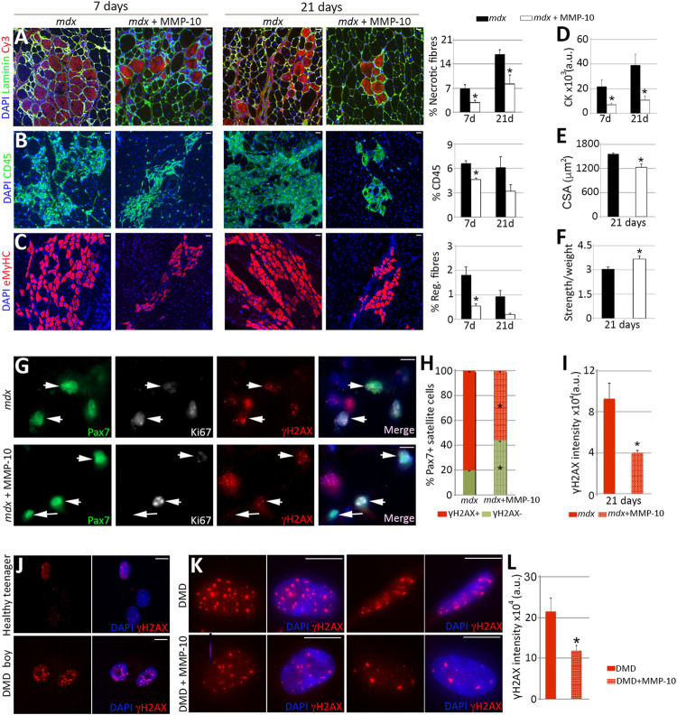
FIGURE 7.
MMP-10 improves dystrophic phenotypes. (A–I) Representative images of gastrocnemius muscles from control and MMP-10-treated mdx mice immunostained by using antibodies to the proteins indicated 7 and 21 days after treatment administration. Necrotic fibers (A), immune cells (B) and new regenerating fibers (C) localized in muscles of control and treated mdx mice after MMP-10 delivery. CK serum levels (D), CSA (E) and muscle strength (F) in the experimental groups. TA muscles of control and MMP-10-treated mdx mice co-immunostained for Pax7, Ki67 and γH2AX (G). Arrowheads identify Pax7+γH2AX+ satellite cells, while arrows point Pax7+Ki67−γH2AX− satellite cells. Percentage of Pax7+γH2AX+ satellite cells (H) and average γH2AX accumulation in Pax7+γH2AX+ satellite cells (I) analysed in muscles from control and MMP-10-treated mdx mice after 21 days. Bars represent the mean ± SEM of at least three animals per condition. (J–L) Representative images of myogenic cells derived from a 19 years-old healthy boy and a 5 years-old Duchenne patient stained for γH2AX (J). Human dystrophic muscle cells stained for γH2AX after 8 days in culture with or without MMP-10 (K). γH2AX intensity accumulation (L) in treated and untreated human cells. Scale bar: 40 μm (A–C, G), 5 μm (J, K). Bars in L show the mean ± SEM of four independent experiments carried out from same human cells. * designates significance between experimental groups, where * p < 0.05. CK, Creatine Kinase; CSA, Cross Sectional Area; a.u, arbitrary units; DMD, Duchenne muscular dystrophy.

