
Fig 1.
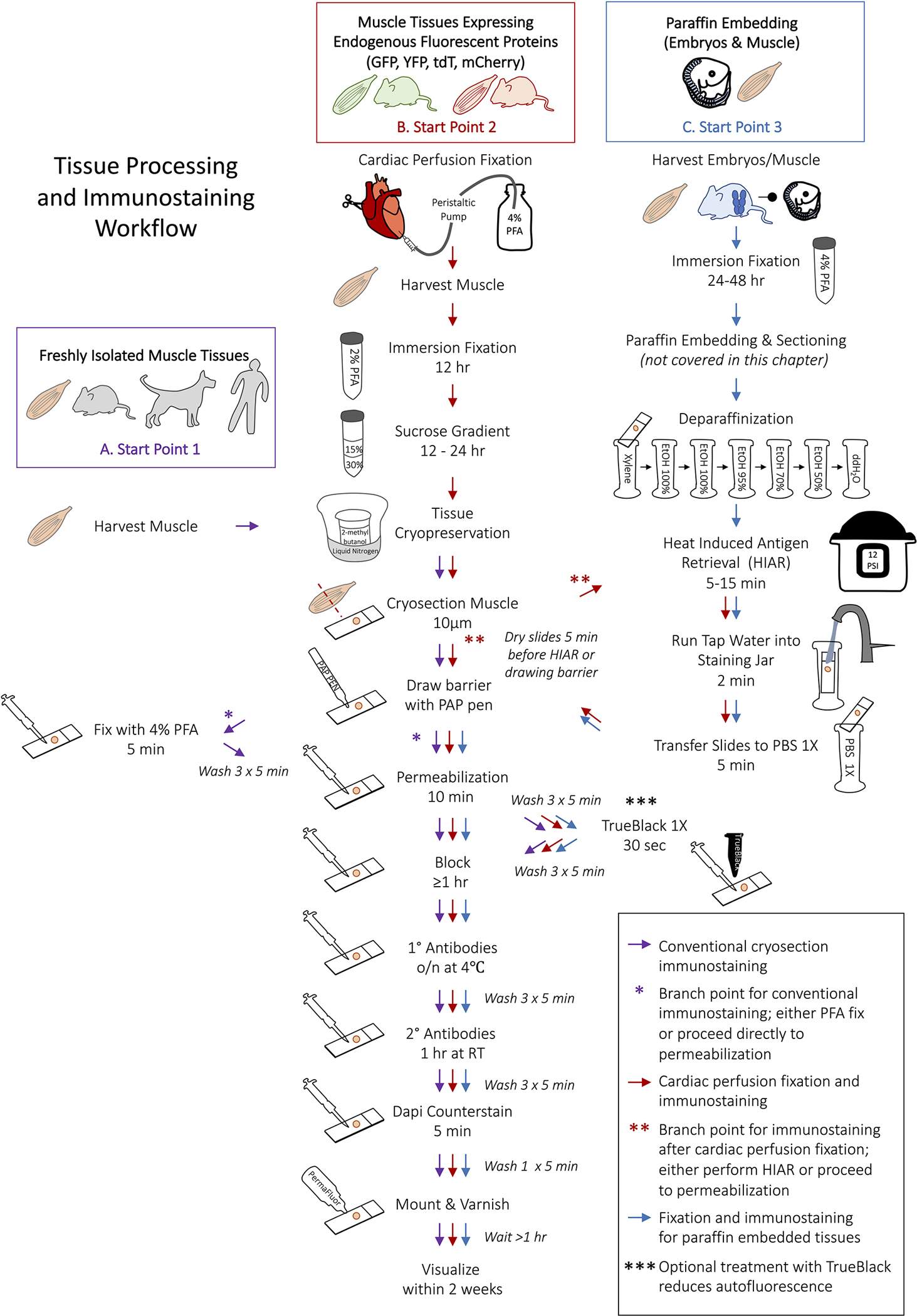
Schematic representation of the workflow for myogenic tissue processing and immunofluorescence staining. (A) Begin from Start Point 1 to process fresh tissue for immunostaining (purple arrows). After surrounding tissues with the hydrophobic barrier, PFA-fixation can be omitted for antigens that are highly sensitive to formaldehyde (purple asterisk). (B) The processing and immunostaining of muscle that requires cardiac perfusion fixation begins at Start Point 2 (red arrows). Heat Induced Antigen Retrieval (HIAR) is not always required for perfusion fixed tissues but is necessary for some primary antibodies (red asterisks). (C) Select Start Point 3 for fixing tissues prior to paraffin embedding and for the subsequent immunostaining of paraffin embedded tissue sections. HIAR is almost always recommended when immunostaining paraffin embedding tissues. Treating tissues with TrueBlack is optional but particularly useful for quenching autofluorescence in the green channel (triple asterisks). Start points A-C correspond to Table 1.