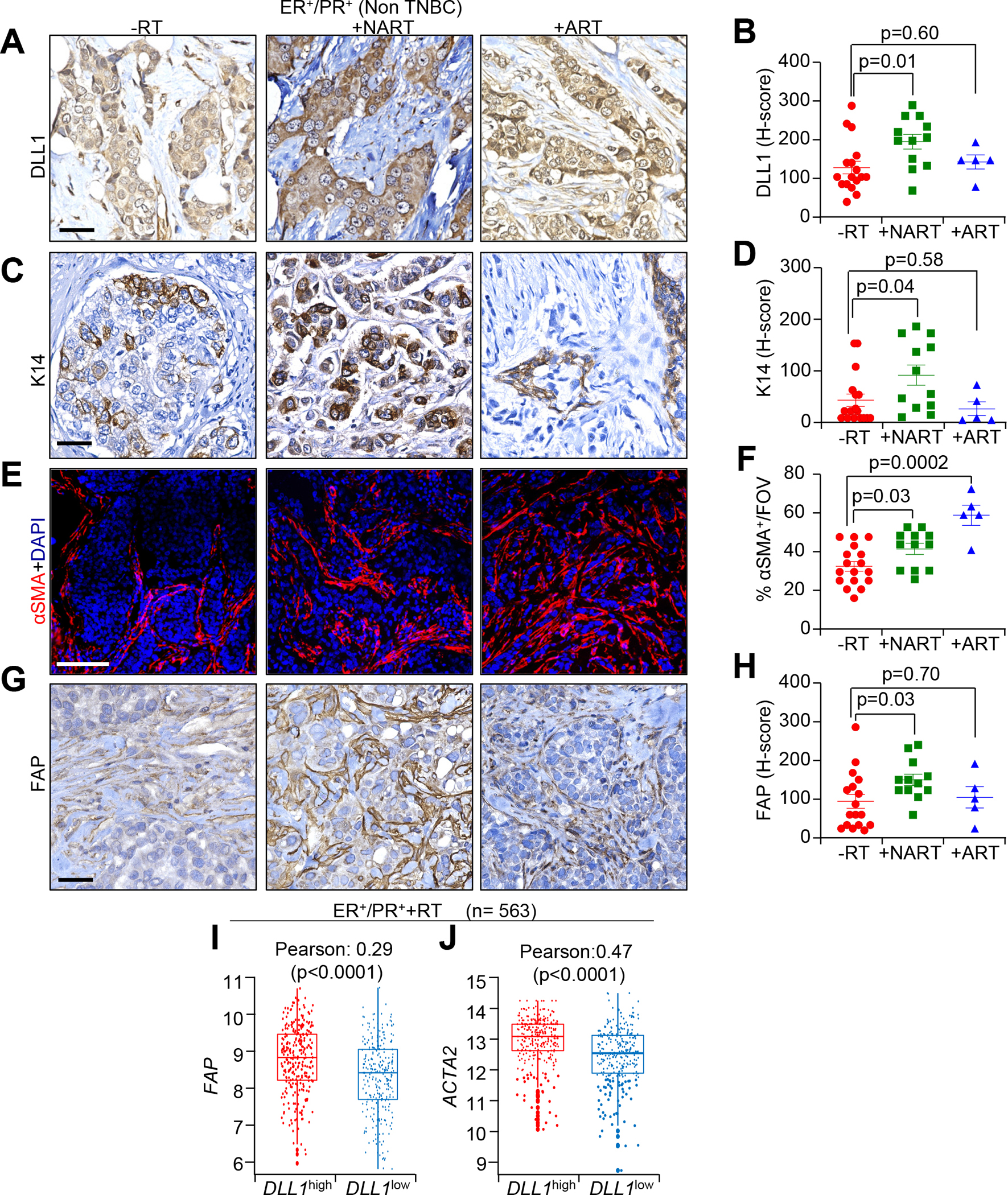
Figure 1.

Radiation increases the expression of DLL1 in tumor cells and FAP+ and αSMA+ CAFs in ER+/PR+ luminal breast cancer patients following RT. A-D, IHC images and H-score quantification of DLL1 (A and B) and K14 (C and D), E, IF images and F, quantification of αSMA G, IHC images and H, H-score quantification of FAP in radiation untreated (-RT), neoadjuvant radiation treated (NART) and adjuvant radiation treated (ART) luminal patient tumors. The dots in each scatter plot represent each patient tumor/group. (n=17 for -RT; n=12 +NART and n=5 +ART tumors). I and J, METABRIC dataset demonstrates correlation between DLL1 (I) and ACTA2 (J) expression in +RT ER+/PR+ (n=563) luminal breast cancer patients. Data are presented as the mean ± SEM. FOV stands for field of view. Unpaired student’s t test was used to calculate p values. Scale bars, 100 μM.
