
Figure 2.
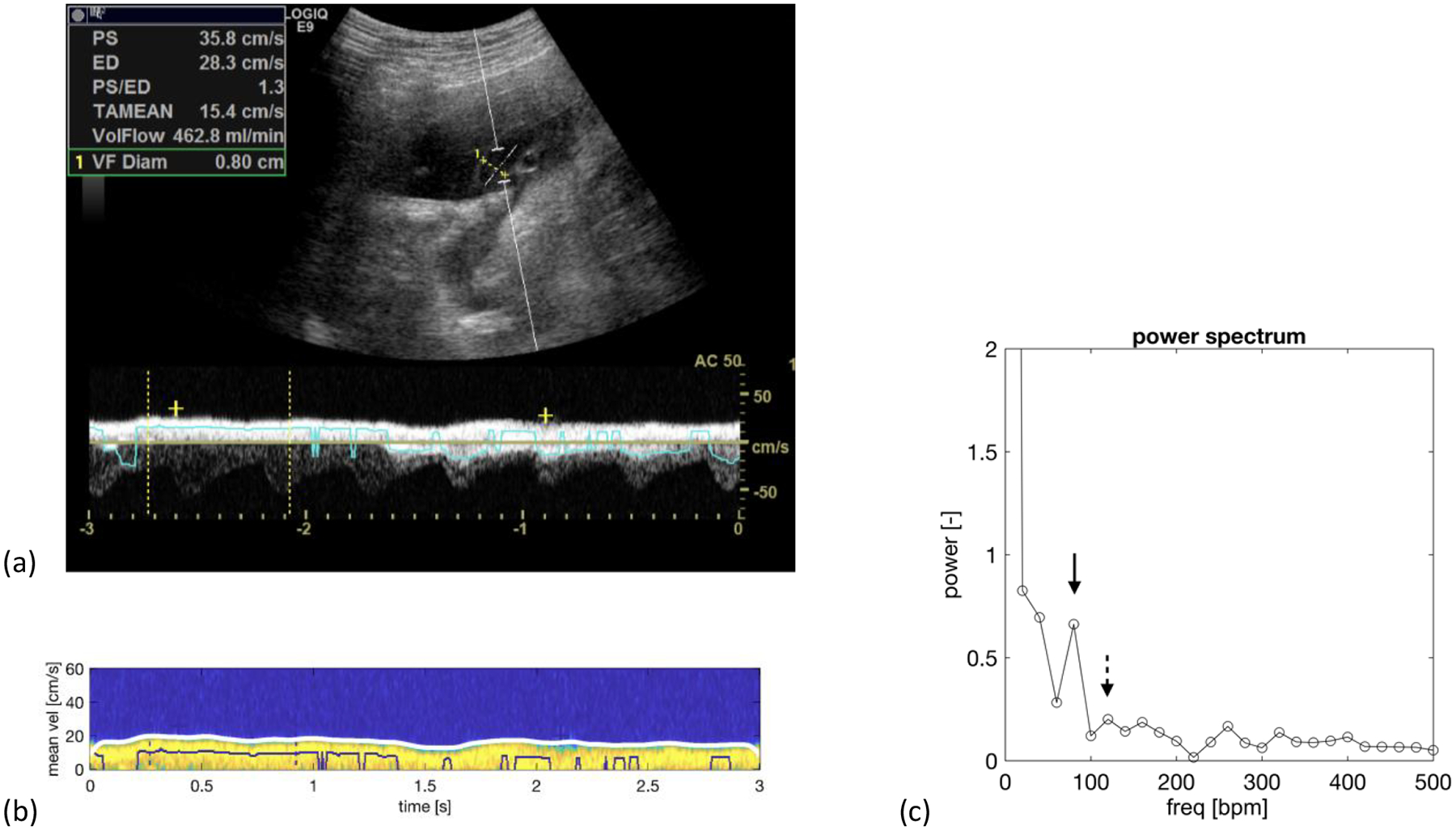
Normal fetus at 34-week 5-day. a) Duplex spectral Doppler of the umbilical vein that has been angle corrected to 50 degrees. b) A colorized version of the venous spectrum with the envelope trace along the upper edge of the spectrum in white. c) Power spectrum showing a maternal peak (solid arrow) at 80 beats/min with a spectral power of 0.663, which is larger than the fetal peak at 120 beats/min with a spectral power of 0.201 (dashed arrow). Note: The zero-frequency peak (DC) is not shown to improve the visibility of the maternal and fetal spectral components. ‘bpm’ equals beats per minute.