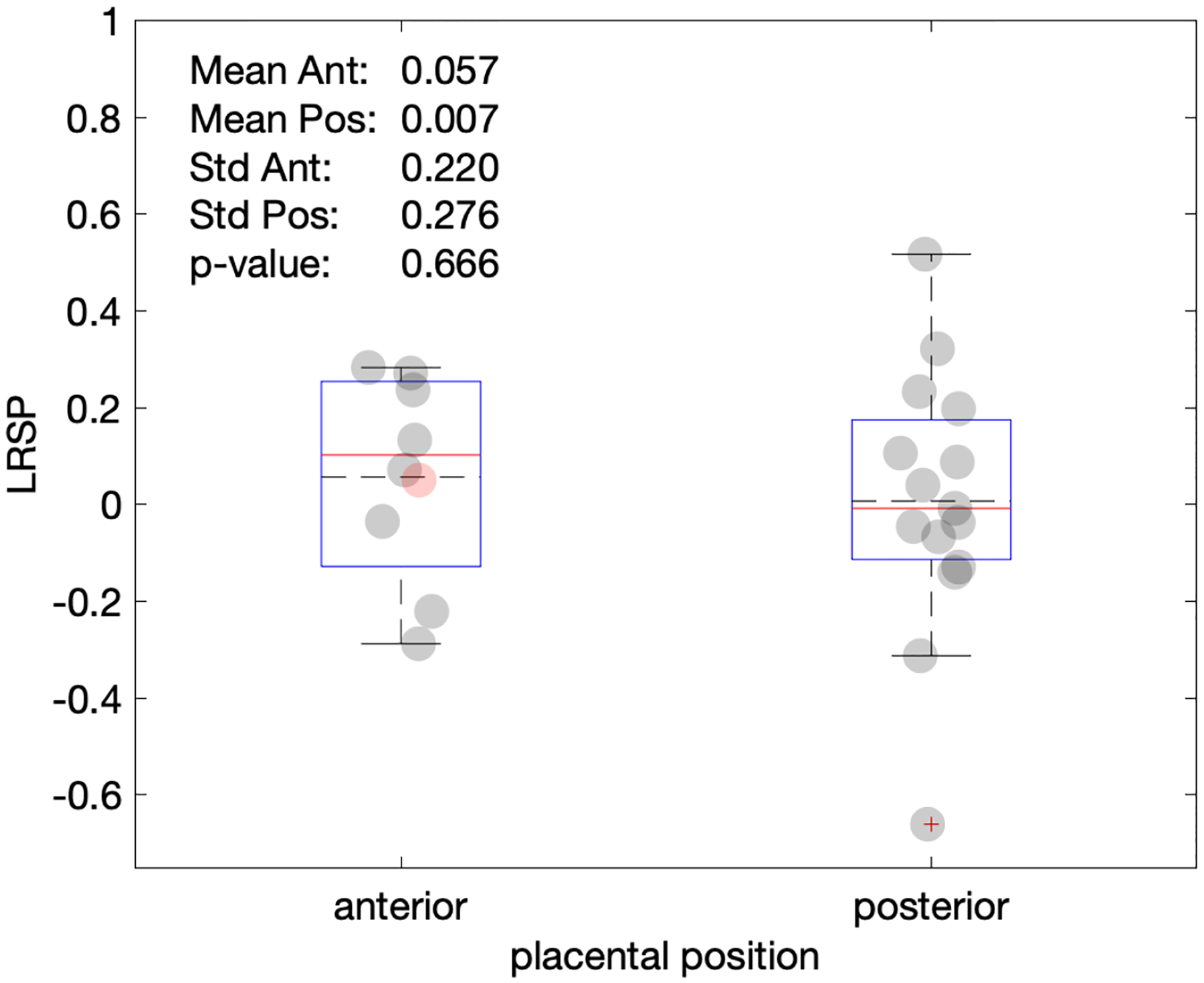
Figure 6.

Plot of LSRP vs. placental position. Most cases investigated here were either anterior or posterior. Only one fundal position (red marker) was observed, which is here arbitrarily placed with the anterior group, but neither included in the box statistics nor in the p-value shown.
