
Figure 8.
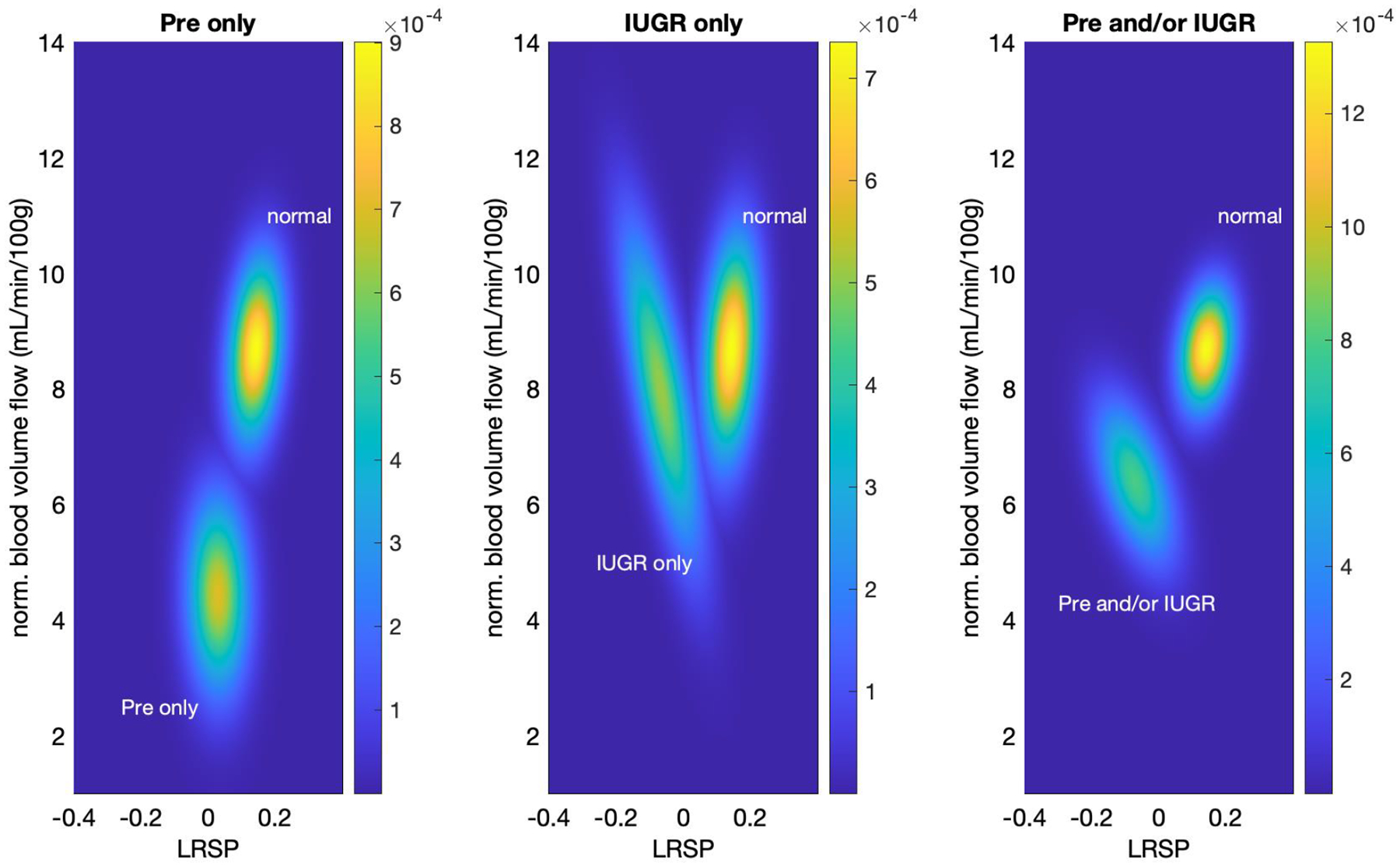
Two-dimensional classification of pathological and normal cases. P-values were computed based on a linear regression model ANOVA analysis. PW Doppler (LRSP) and blood volume flow measurements are the independent variables for the linear regression model with classifications of normal pathology, preeclampsia, IUGR or either/both being the dependent variables. Left: Pre-eclampsia cases predominantly differ from normal with respect to normalized blood volume flow. Middle: IUGR cases predominantly differ from normal with respect to LRSP. Right: The combined cohort of pre-eclampsia and/or IUGR cases shows a combined effect of LRSP and normalized blood volume flow with significance in both LRSP only as well as with normalized blood volume flow (see Table 1).