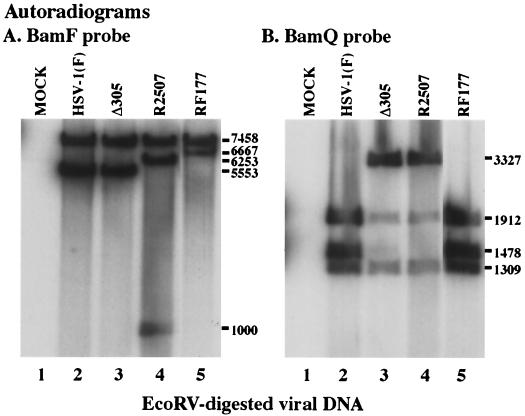
FIG. 3.
Autoradiographic images of Southern hybridizations using the BamF (A) and BamQ (B) probes. EcoRV-digested DNA from mock- (lane 1), HSV-1(F)- (lane 2), Δ305- (lane 3), R2507- (lane 4), or RF177- (lane 5) infected V49 cells was separated in a 0.8% agarose gel and probed with the BamF fragment, containing the UL49 gene, or the BamQ fragment, containing the UL23 gene, as described in Materials and Methods. The size of EcoRV fragments hybridizing to BamF or BamQ probes appear to the right as molecule weights. DNA from several independent RF177 plaques was analyzed, and lane 5 shows the result for the virus isolate used throughout our studies.

