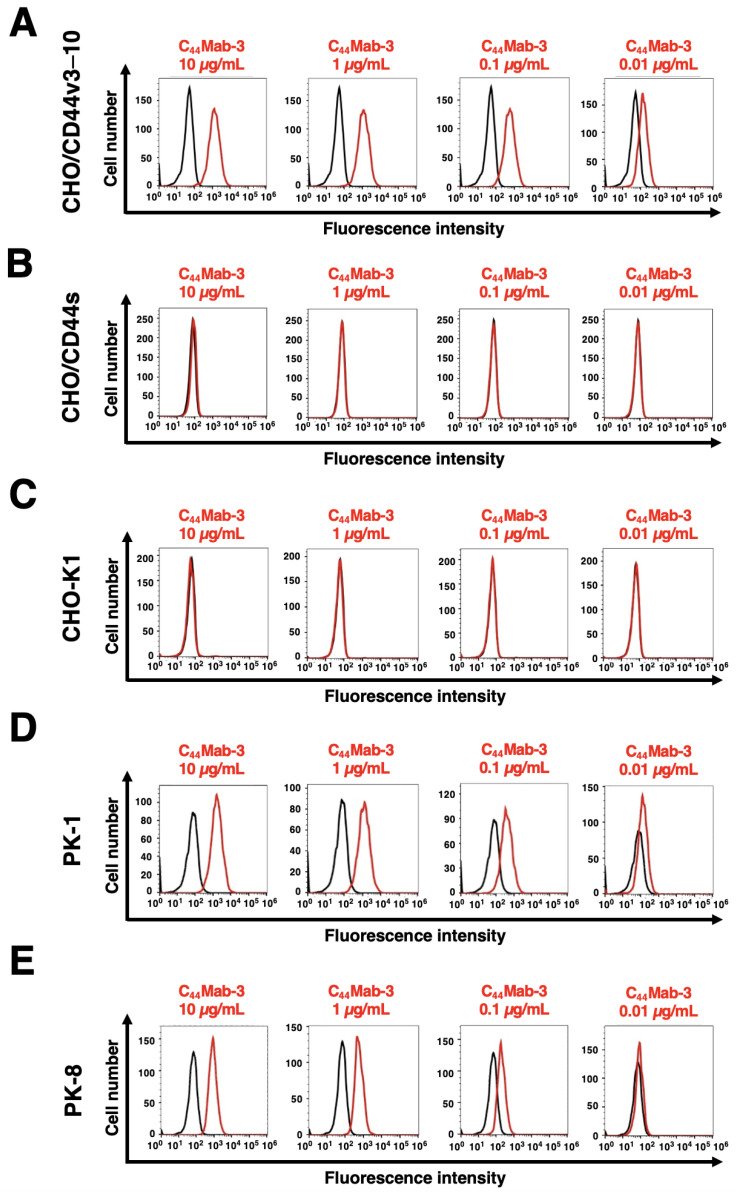
Figure 2.
Flow cytometry using C44Mab-3 against CD44-expressing cells. CHO/CD44v3–10 (A), CHO/CD44s (B), CHO-K1 (C), PK-1 (D), and PK-8 (E) cells were treated with 0.01–10 µg/mL of C44Mab-3, followed by treatment with Alexa Fluor 488-conjugated anti-mouse IgG (Red line). The black line represents the negative control (blocking buffer).

