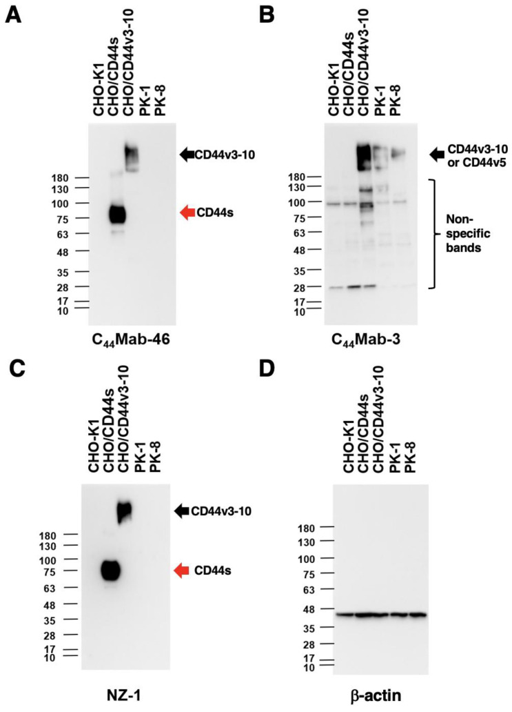
Figure 4.
Western blot analysis using C44Mab-3. The cell lysates of CHO-K1, CHO/CD44s, CHO/CD44v3–10, PK-1, and PK-8 (10 µg) were electrophoresed and transferred onto polyvinylidene fluoride (PVDF) membranes. The membranes were incubated with 10 µg/mL of C44Mab-46 (A), 10 µg/mL of C44Mab-3 (B), 0.5 µg/mL of an anti-PA16 tag mAb (NZ-1) (C), and 1 µg/mL of an anti-β-actin mAb (D). Then, the membranes were incubated with anti-mouse immunoglobulins conjugated with peroxidase for C44Mab-46, C44Mab-3, and anti-β-actin. Anti-rat immunoglobulins conjugated with peroxidase were used for NZ-1. The red arrows indicate CD44s (~75 kDa). The black arrows indicate CD44v3–10 or CD44v5 (>180 kDa).

