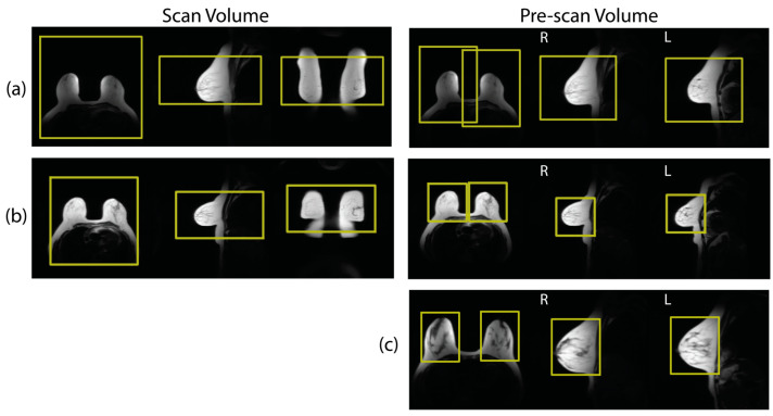
Figure 1.
Examples (a,b) show scan volumes and pre-scan volumes as prescribed during clinical breast MRI. Both (a,b) come from the same patient but from exams on different days. Note that the placement of scan volumes was relatively consistent across both exams. However, the pre-scan volume placements were dramatically different between exams. Example (c) shows an example from a different patient with offline volumes generated by expert users that was used to train pre-scan volume placement.

