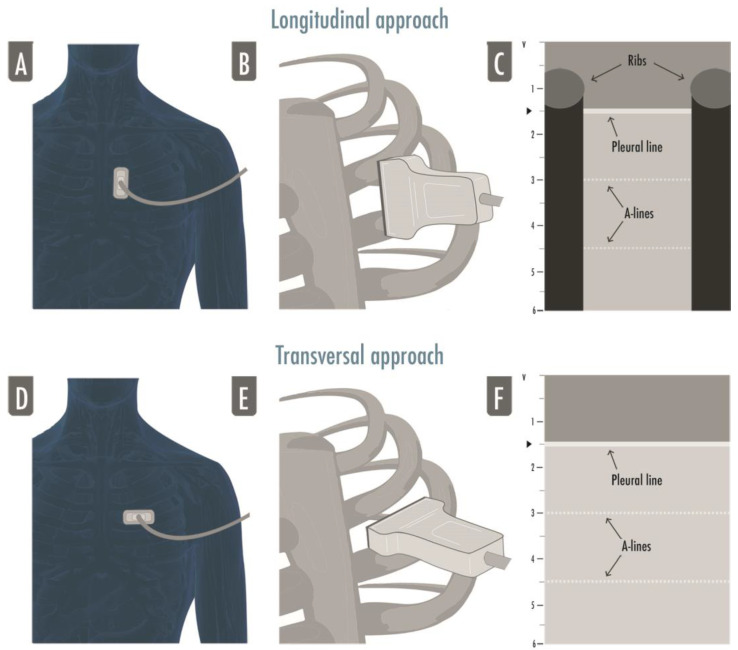
Figure 1.
The figure illustrates both approaches to lung ultrasound: the longitudinal approach (A–C) and the transversal approach (D–F). In the longitudinal approach, the probe is aligned to the craniocaudal axis of the patient and perpendicular to the ribs’ axis (A,B), giving the characteristic ultrasonographic image of the two ribs and their shadows defining the pleural line in the middle, the so-called bat sign (C); in the transversal approach the probe is placed in the intercostal spaces, parallel to the ribs’ axis (D,F), so that a larger pleural section can be displayed without any rib’s shadows visualized (F).

