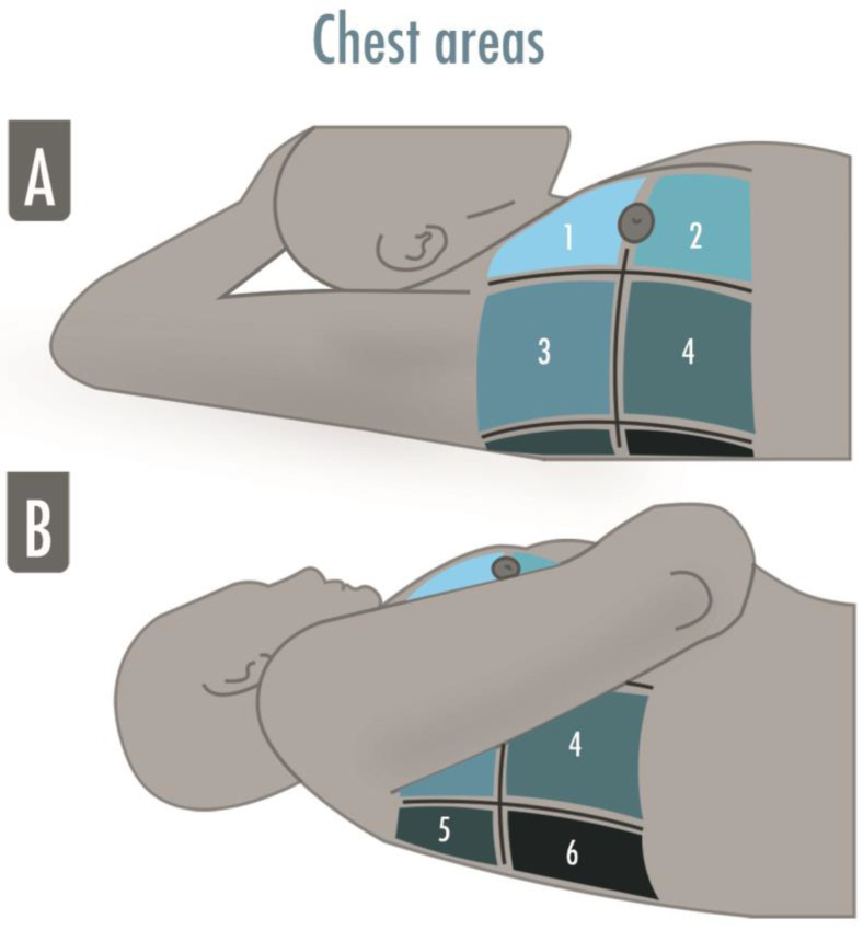
Figure 2.
The illustration shows the six-areas division of a hemithorax in a supine patient for a comprehensive lung ultrasound examination. The anterior and the posterior axillary lines divide the hemithorax into three parts, which are finally divided into superior and inferior. The areas may be numbered from “1” to “6”, corresponding, respectively, to the anterior–superior and the inferior–posterior areas. (A) Zone 1 and 2 are superior and inferior anterior scans, zone 3 and 4 are superior and inferior lateral scans; (B) Zone 5 and 6 are superior and inferior posterior scans.

