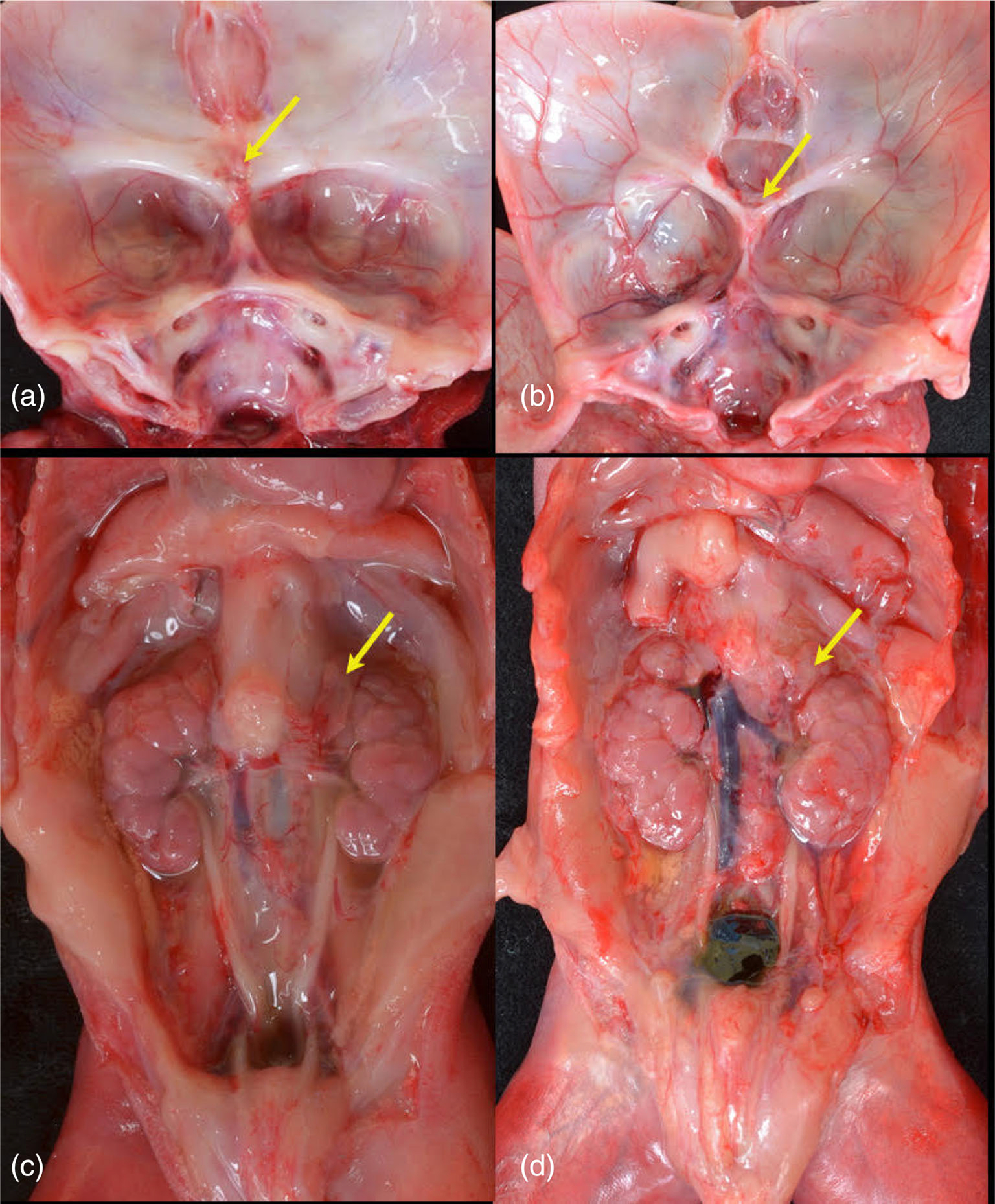
FIGURE 5.

Internal structures. Comparison of skull bases of Proband 3 (a) and Proband 4 (b). Proband 3 (a) with absent sella turcica. Arrow indicates fused optic nerve. No pituitary was present, even on subserial sectioning of the sphenoid. Proband 4 (b) demonstrates wide, shallow sella anterior to (arrow) fused optic nerve. A small pituitary was present. In both fetuses, the hypothalamus was disorganized and no infundibulum was present. Retroperitoneum demonstrating small adrenal glands (arrows) in Proband 3 (c) and Proband 4 (d).
