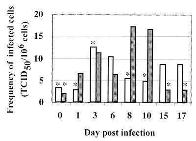
FIG. 3.
MVV is associated with dendritic cells in afferent lymph. Afferent lymph dendritic cells from a sheep infected with MVV subsequent to cannulation were purified by metrizamide gradient centrifugation followed by FACS sorting. CD14− CD1blo and CD14lo CD1bhi dendritic cells were analyzed for the presence of infectious virus by cocultivation with indicator skin cells. The frequency of infected cells is shown versus time postinfection. Open histogram, CD14− CD1blo dendritic cells; closed histogram, CD14lo CD1bhi dendritic cells; ∗, no detectable virus, but bar indicates sensitivity of assay, i.e., maximum level of infectivity which theoretically could have been present but undetected. Day 0 samples were taken before infection. Mean purity of FACS-separated cells was 91% (range of 67.1 to 97%) after adjustments for autofluorescence and spectral overlap.

