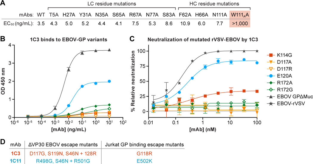
Figure 4. Binding and neutralization analysis of monoclonal antibodies (mAbs) 1C3.The IMGT numbering scheme is used.
(A) Amino-acid residue changes in 1C3. Only one change in 1C3, W111A, compromised tight binding of Ebola virus (EBOV) glycoprotein (GP). All other single amino-acid residue changes could be accommodated, presumably by other key contacts remaining. (B and C) Changes in GP residues. (B) Point changes in GP at 3 of 4 key residues result in reduced binding by 1C3, presumably because these single amino acids form more than one contact point each in the tripartite epitope. (C) Point changes at all four key residues result in reduced neutralization of recombinant vesicular stomatitis virus expressing EBOV GP (rVSV-EBOV) by 1C3. (D) Viral escape mutants from 1C3 and 1C11. Left column: Mutations identified in plaque-purified ΔVP30 Ebola virus isolates grown in the presence of 1C3 or 1C11. Right column: Mutations identified in Jurkat cell lines expressing randomly mutated EBOV GP that were selected for loss of binding to 1C3 or 1C11 by FACS sorting.

