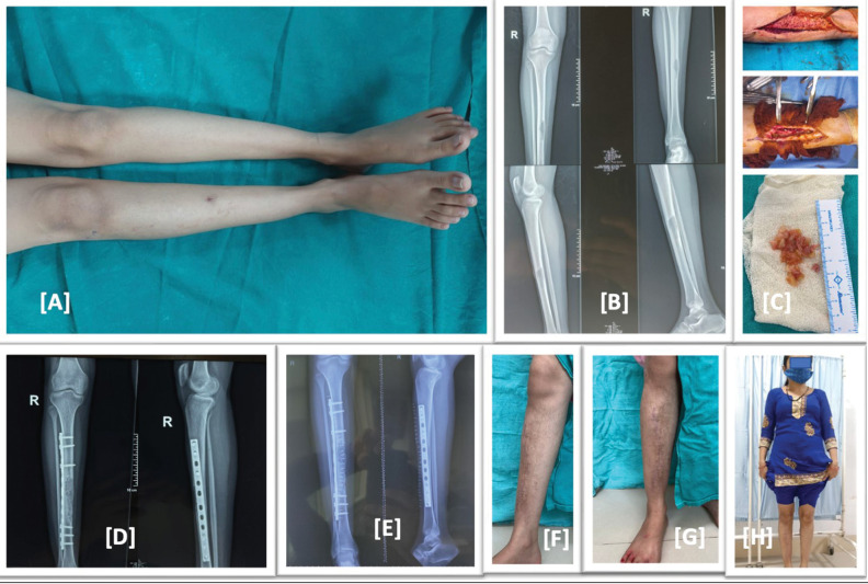
Figure 1.
(A) Preoperative image, right leg with scar mark of biopsy and mild swelling. (B) Preoperative X-ray showing expansile lytic lesion in mid shaft tibia. (C) Intraoperative images, excised hydatid cyst mass. (D) Immediate postoperative X-ray showing in corporation of allograft and fixation by anterolateral tibial plate. (E) Post operative X-ray at 6 month follow up showing well incorporation of allograft. (F–H) Clinical images at 6 month follow up showing healed scar mark no swelling or deformity and full weight bearing of the patient with full return to activity of daily living

