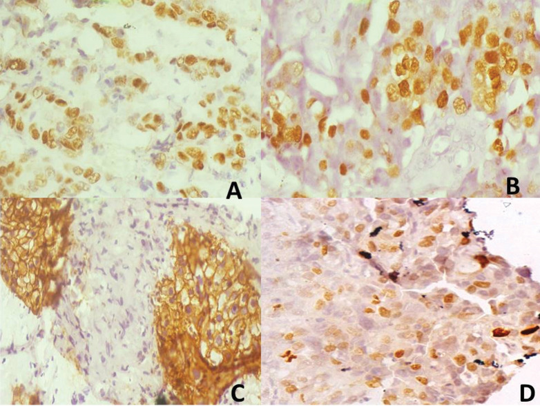
Figure 1.
Composite photomicrographs of (a) (×400): Representative oestrogen receptor (ER) positive tumour (3+). (b) (×400): Representative progesterone receptor (PR) positive tumour (3+). (c) (×400): Representative human epidermal receptor-2 (HER-2) positive tumour (3+). (d) (×400): Representative Ki-67 positive tumour, proliferative index about 35%

