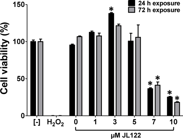
Fig. 2. EPC cell viability at 24 and 48 h post-exposure to JL122.
EPC cells were exposed to up to 10 μM JL122, vehicle control only (0.1% DMSO; 0 μM JL122), or hydrogen peroxide (40 mM final concentration) for 24 or 72 h in the presence of light. Formazan dye absorbance was measured at 450 nm and used to assess % cell viability. Negative control [-] = no compound. DMSO concentration varied for each JL122 dose depending on dilution: 10 μM JL122 had 0.1% DMSO; 1 μM JL122 had 0.01% DMSO. Cytotoxicity was absent when JL122 concentrations were ≤ 5 μM. Toxicity to cells was present when exposed to ≥7 μM JL122. A hyperplastic response occurred with 3 μM JL122 at 24 h post-exposure. Data represents mean cell viability ± SE (n=5; triplicate samples are shown and a similar trend was observed in 5 repeated experiments) when normalized to no treatment (negative control). *: P<0.001, ANOVA, Tukey’s Multiple Comparison Test.

