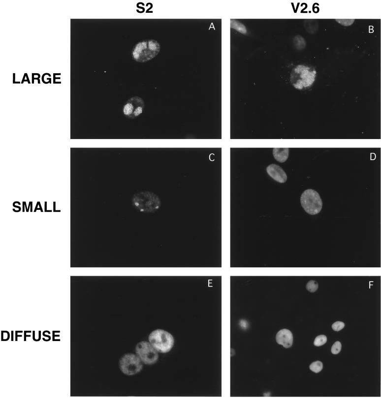
FIG. 3.
Examples of different patterns of intranuclear localization of replication proteins in S2 and V2.6 cells infected with HSV-1 KOS strain. Cells were stained with the ICP8 antiserum 3-83. S2 cells expressing wild-type ICP8 (left) and V2.6 cells expressing d105 ICP8 (right) were used. Examples of large compartments (A and B), small compartments (C and D), and diffuse staining (E and F) are depicted.

