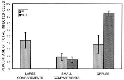
FIG. 4.
Percentages of infected cells in each class of intranuclear localization of replication proteins. Cells were stained by immunofluorescence for ICP8 with 3-83 rabbit serum, and infected cells were counted and classified as containing large compartments, small compartments, or diffuse staining. The percentage of each in S2 and V2.6 cells are shown. More than 400 cells were counted for each cell line in three separate experiments with at least two coverslips of infected cells per sample per experiment. The error bars show standard deviations of the results of three experiments.

