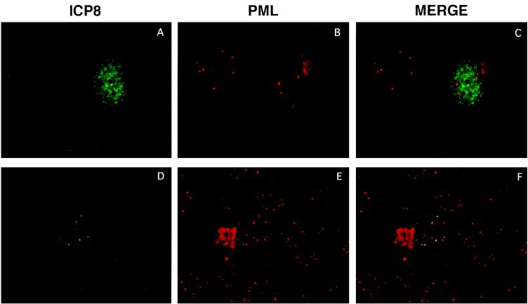
FIG. 9.
Dual staining of ICP8 and PML in C105 cells infected with the ICP0 null mutant virus n212. A representative cell that formed small replication compartments dually stained with antibodies against ICP8 (39S) and PML. The yellow staining in the merged image indicates colocalization of the two proteins.

