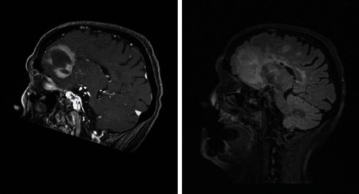
Figure 4.

Extensive white matter changes with multiple patchy areas of intense enhancement in the left anterior and inferior frontal lobes. Radiographic differential diagnosis: PCNSL, high-grade primary neoplasm, leukoencephalopathy, and primary leukoencephalopathy with immune-reconstitution inflammatory syndrome.
