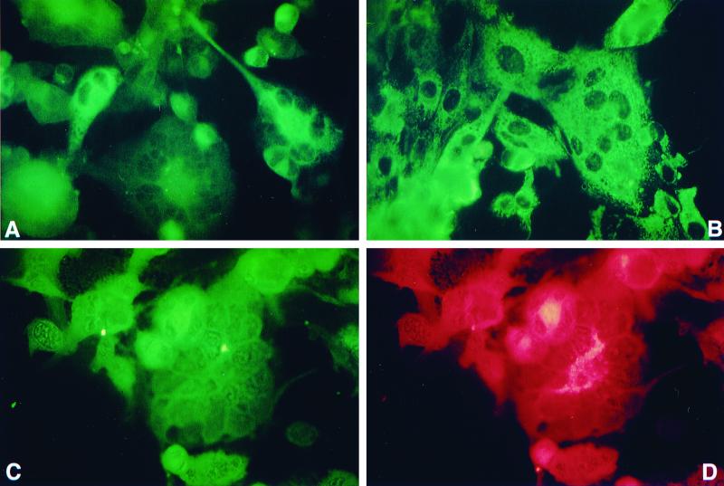
FIG. 8.
Detection of serotype 1 MDV-specific pp38 in QT35 cells inoculated with HVT-infected CEF (QT35/HVT). (A) QT35/HVT cells stained with pp38-specific MAB H19 and FITC-conjugated goat anti-mouse immunoglobulin (heavy and light chains) [Ig (H+L)]. Note the cytoplasmic fluorescence. QT35 cells or QT35 cells inoculated with CEF and treated in a similar way were negative (data not shown). (B) Positive control for pp38 staining. CKC infected with the JM-16 strain of MDV were stained as described for panel A. (C and D) QT35/HVT cells were examined for coexpression of serotype 1 MDV pp38 and an HVT glycoprotein using MAbs H19 and L78, respectively, FITC-conjugated goat anti-mouse Ig (H+L), and TRITC-conjugated goat affinity-purified anti-mouse IgG. Cells were examined for expression of pp38 and HVT glycoprotein by switching filters appropriate for exciting FITC and TRITC, respectively.

