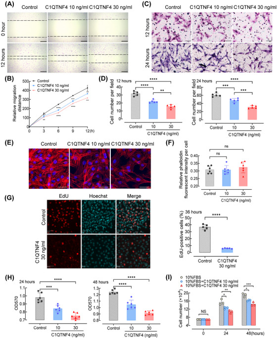
FIGURE 1.

Recombinant C1QTNF4 protein suppresses the migration and proliferation of VSMCs. (A) Confluent VSMC monolayers were treated with or without 10 and 30 ng/mL recombinant C1QTNF4 protein for 24 h before scratch wounding. The cells were kept in culture for an additional 3, 6, 9 and 12 h before analysis. Representative images of the migration assays at 0 h and after 12 h of culture. The dotted line indicates the wound edge. Scale bar, 200 μm. (B) The mean migration distance of VSMCs was quantified at 3, 6, 9 and 12 h after scratching. ± SEM, n = 7. (C) VSMCs were seeded on the transwell dishes, exposed to C1QTNF4 10 ng/mL, 30 ng/mL or control for 12 or 24 h, and then subjected to transwell migration assay. Scale bar, 50 μm. (D) The cell number per field was counted in the transwell assay (± SEM; n = 5). (E) Representative confocal image of 3 independent experiments showing the effect of recombinant C1QTNF4 protein (10 or 30 ng/mL) on VSMC stress fibres at 48 h. Scale bar, 20 μm. (F) Quantification of relative phalloidin fluorescent intensity per cell with different C1QTNF4 concentrations (± SEM; n = 6). (G) Representative images of EdU incorporation in VSMCs incubated with or without 30 ng/mL recombinant C1QTNF4 protein for 36 h. Nuclei were stained with Hoechst 33342 (blue), and proliferating cells were labelled with EdU (red). Scale bars, 50 μm. Percentages of EdU‐positive cells were calculated. ± SEM, n = 5 per group. (H) The MTT assay was used to analyse the number of live cells cultured in different concentrations of C1QTNF4 at 24 and 48 h (± SEM; n = 6). (I) Cell counting assays were performed with different concentrations of C1QTNF4 (± SEM; n = 3). *p < .05, **p < .01, ***p < .001, ****p < .0001; NS indicates no significance.
